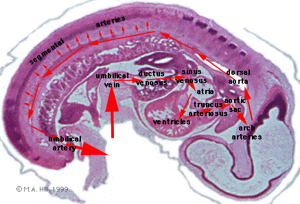
|
E2,E6:
Dorsal branches of single dorsal aorta.
F5:
Dorsal aorta giving rise to a ventral branch
(mesenteric artery), and branch to mesonephros (the
developing kidney).
F7:
Dorsal aorta sending branch into mesonephros (to
glomerulus).
G2,G3:
Dorsal aorta curving around flexed trunk of embryo.
Note spinal cord cut twice.
G4,G5:
Sections gradually passing dorsally beyond
curvature of single dorsal aorta.
Return to G4. We now will follow the aorta in
the lumbar region of the embryo as it passes into
the sacral region.
G2,G1:
Single dorsal aorta ventral to sacral (smaller)
spinal cord.
F7-E7:
Bifurcation into paired dorsal aortae.
E6,E5:
Lateral major branch of dorsal aorta becomes
umbilical artery: it reaches a crest in E5 (i.e.
not seen in E4) and then "descends" in the ventral
body wall to F2, where it enters the body stalk. We
will follow this complex twisting again when we
study the development of the hindgut.
|