UNSW Embryology
|

|
DEVELOPMENT OF THE HEART AND
CARDIOVASCULAR SYSTEM
|
Embryology Home
Page
|
|
|

|
|
Page
Links ventricles
into the aortic systemAbout
Notes ventricles
into the aortic systemAbout
Notes
Next page caudal
path of the arterial blood
|
Page
1 | Introduction
Page
2 | Questions
| Abnormal
| OMIM
| Refs
Page
3 | Pig
Stage 13/14
Page
4 | Human
(Stage22)
Page
5 | Selected
Human highpower
Text only
page | WWW
Links
|
|
|
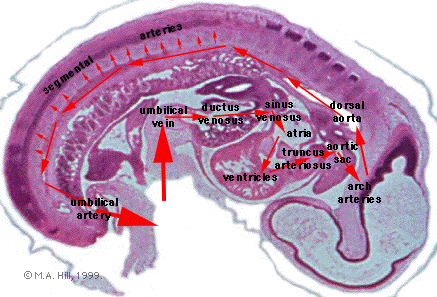
G7
An overview of blood flow through the embryo of
oxygenated blood. Note the umbilical artery and
veins anastomose within the chorion of the
placenta, there is no direct connection of maternal
and foetal blood.
Maternal Blood
umbilical vein
liver
anastomosis
ductus venosus
sinus venosus
atria ventricles
truncus arteriosus
aortic sac
aortic arches
dorsal aorta
pair of umbilical arteries
Maternal Blood
|
|
High pressure
pathway from the ventricles into the aortic
system.
|
|

|
D7:
Apex of left
ventricle: spongy network of endocardium; a small
dorsal cavity (part of the intraembryonic coelom
which will become the pericardial cavity - no
pericardium yet); mesenchymal jelly of body
wall.
|
|

|

|
D6,D5:
Tip
of right ventricle; left ventricle; I-E
coelom; liver, with its ventral transverse
border, the
septum
transversum.
|
|
|

|

|

|
|
D2:
Trabeculae of L and R ventricles;
interventricular septum; interembryonic
coelom; sinus
venosus caudal tip of part of left atrium
= L auricular appendage.
|
Dl:
Endocardial jelly at dorsal wall of
interventricular septum.
|
C7:
Ventricles forming
interventricular
septum (muscular portion); dorsal
endocardial jelly communication of
L auricle and
L atrium; R atrium;
R auricle.
|
|
|

|
C6:
Aperture in interatrial septum, the
ostium
(foramen) primum. L and R atrioventricular canals.
Note the L auricle is not separated from L atrium,
the plane of section has caught the wall fold at
the region where the auricle connects.
|
|

|
C5:
Communication of L auricle with left atrium.
|
|

|
C4:
Transition from R ventricle to the outflow tract,
including the
truncus arteriosus
and complete
interatrial septum
(cf. C5).
|
|

|
C3:
Truncus
arteriosus. Atria. Right venous valve in R
atrium.
C7-C3:
Return to C7 and proceed to C3, noting how left
ventricular blood has to pass obliquely across
right ventricle to exit from heart via the outflow
tract and truncus arteriosus.

|
|

|

|
C2,
C1
Truncus
arteriosus shifting to midline.
Jelly and
mantle of the truncus.
Cranial end of
R venous
valve. Note also extent of I-E coelom;
thin body wall.
|
|
|

|

|

|
B7-B5:
Attachment of truncus arteriosus to
ventral
body wall and to dorsal roof of intra
embryonic coelom. Note the primordium of
the
transverse
pericardial sinus in B6,
caudal to the attachment of the
truncus.
|
|
|
|

|
B5-B4:
Entry of truncus arteriosus into
aortic sac,
completely embedded in pharyngeal arch
mesectoderm. Note position of aortic sac
in relation to pharynx
and pharyngeal arches.
4th
pharyngeal arch artery on left.
|
|
|

|
Return to B5
and note the small "6th" pharyngeal arch artery
either side of the laterally-compressed
pharynx.
|
|
|
B3:
Cranial end of aortic sac. 4th pharyngeal arch
arteries and emerging 3rd pharyngeal arch artery.
Note dark thyroid primordium ventral to origin of
3rd arch arteries.
|
|
|
B2:
(lst and 2nd pharyngeal arch arteries not seen. 3rd
arch arteries. Bilaterally, communication of 4th
arch artery (at sides of pharynx) with dorsal
aorta.
|
|
|
B1:
3rd arch arteries. Dorsal aortae. (Superior
cardinal veins lateral to aortae).
|
|
|
A7:
On left side, communication of 3rd arch artery with
aorta (i.e. occurring cranial to the 4th arch
communication). From here on the arterial blood is
distributed through fine branches to vessels
outside the brain (pial plexus).
|
|
Next- caudal
path of the arterial blood
|
|
|
|
Next page for
embryonic blood flow
|
caudal path
of the arterial blood
|
About Notes
- Lecture notes from the Anat 3311 1997
Science Embryology course compiled and written by Dr
Mark Hill. Some notes derived from historic
class notes.
- Note Links to OMIM Entries are copies of originals
for computers without internet access. Computers with
internet access can directly access the database.
|
|
|
m.hill@unsw.edu.au
Date Last Modified: 11/3/99
This site maintained by Dr M. Hill
|

|