UNSW Embryology
|

|
RESPIRATORY
SYSTEM
|
Embryology Home
Page
|
|
|

|
|
Page
Links IntroductionReading
Computer
Activities
ObjectivesLearning
activities Development
Overview Pig
OverviewTermsReferencesAbout
Notes IntroductionReading
Computer
Activities
ObjectivesLearning
activities Development
Overview Pig
OverviewTermsReferencesAbout
Notes
|
Page
2 | Abnormalities
| OMIM
| Self
Assessment Questions |
Medline
Page
3 | Pig
Stage 13/14
Page
4 | Human
(St 22) Trachea |
Human (st
22) Lungs
Page
5 | Selected
Human highpower
Text only
page | WWW
Links
|
Introduction
|
Reading
- Human Embryology (2nd ed.) Larson
Ch6: p127-149
- The Developing Human: Clinically Oriented
Embryology (6th ed.) Moore and Persaud Ch11:
p226-233
- Before we Are Born (5th ed.) Moore and
Persaud Ch12; p241-254
- Essentials of Human Embryology Larson Ch6
p81-96
- Human Embryology Fitzgerald and Fitzgerald
Ch18 p113-118
- Additional References- Selected,
Lung
Development, Respiratory
Development
- Search PubMed-
Medline
|
Computer
Activities
UNSW
Embryology:
|
|
|
Embryo Images
Unit:
|
Unit: Body Cavities
|
|
Objectives
- Describe the development of the respiratory
system from the endodermal and mesodermal
components.
- Describe the main steps in the development
of the lungs.
- Describe the development of the diaphragm
and thoracic cavities.
- List the respiratory changes before and
after birth.
- Describe the developmental aberrations
responsible for the following malformations:
tracheo - oesophageal fistula (T.O.F);
oesphageal atresia; diaphragmatic hernia; lobar
emphysema.

|
Learning activities
- Summarise the changes in the respiratory
system and the C.V.S. at birth.??
- To discuss the main features of
physiological maturation of the lung and the
importance of this to fetal and newborn
viability.
- Examine the serial sections of human and pig
with reference to the respiratory system.
- Discuss the selected malformations; tracheo
- oesophageal fistula (T.O.F.) diaphragmatic
hernia and lobar emphysema giving special
reference to the developmental aberrations
causing the malformations to the
individuals
|
|
|
|
Development of the
Respiratory System
- Week 4 laryngotracheal groove forms on floor
foregut
- Week 5 left and right lung buds push into
the pericardioperitoneal canals (primordia of
pleural cavity)
- Week 6 descent of heart and lungs into
thorax. Pleuroperitoneal foramen closes.
- Week 7 enlargement of liver stops descent of
heart and lungs.
Diaphragm- 5 elements contribute to the
diaphragm
- septum transversum- central tendon
- 3rd to 5th somite- musculature of
diaphragm
- ventral pleural sac- connective tissue
- mesentry of oesophagus- connective tissue
around oesophasus and IVC
- pleuroperitoneal membranes- connective
tissue around central tendon
Blood Supply-
- pulmonary system not "functional" until
after birth
- 6th aortic arch arteries generate pulmonary
areries
- veins drain into pulmonary vein then left
atrium
- branches from dorsal aorta generate
bronchial arteries
Lung Histology-
- 4 periods
- 5-17 week pseudoglandular
- 16-25 week canalicular
- 24-40 week terminal sac
- late fetal-8years alveolar
- month 3-6 lungs appear glandular
- end month 6 alveolar cells type 2 appear and
begin to secrete surfactant
- month 7 respiratory bronchioles proliferate
and end in alveolar ducts and sacs
|
|
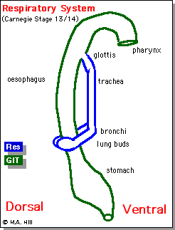
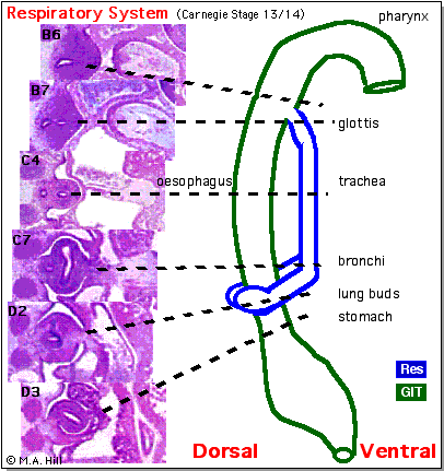
Pig Serial Sections
|
|
|
|
References
- Selected Lists of References from PubMed
March 1999 search results are available for
School of Anatomy computers without internet
access. Computers with internet access can
search from either Page
2 or PubMed
Internet Access
|
|
|
|
Respiratory Terms
- atresia- obstruction.
- eppiglottis- develops from
hypobrachial eminence.
- fistula- abnormal comunication.
- hypopharyngeal eminence- fusion of
3rd pharyngeal arches, precursor of root of
tongue.
- laryngotracheal groove- forms on
anterior (ventral) wall of pharynx, gives rise
to larynx, trachea, respiratory tree.
- larynx- lining from endoderm,
cartilage from pharyngeal arch 4 and 6.
- lung buds- primordia of lungs.
- parietal pleura-outer lining of
pleural cavity derived from epithelia of
pericardioperitoneal canals from intraembryonic
coelom.
- pleural cavity- walls derived from
pericardioperitoneal canals -> intraembryonic
coelom ->coelomic spaces -> lateral
mesoderm -> mesoderm.
- pleuropericardial fold- restricts the
communication between pleural cavity and
pericardiac cavity, contains cardinal vein and
phrenic nerve.
- pleuroperitoneal membrane- forms
inferiorly at transverse septum to separate
peritoneum from pleural cavity.
- septum transversum- mesoderm
separating thoracic cavity and yolk sac, forms
central tendon of diaphragm (and some of
liver?).
- stenosis- narrowing
- surfactant- a detergent secreted by
Type 2 alveolar cells between alveolar
epithelium. Functions to lower surface tension,
allowing lungs to remain inflated.
- visceral pleura- inner lining of
pleural cavity derived from contact epithelia
with lung bud of pericardioperitoneal canals
from intraembryonic coelom.
|
|
About Notes
- These lecture notes from the Embryology course
compiled and written by Dr Mark Hill.
- Note Links to PubMed Medline Entries are copies of
originals for computers without internet access.
Computers with internet access can directly access the
database.
- Note that reference lists are only relevant to the
date that the original search was carried out.
|
|
|
m.hill@unsw.edu.au
Date Last Modified: 11/3/99
This site maintained by Dr M. Hill
|
|