|
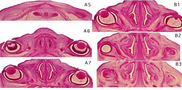
B4:
Tongue. Otic capsule. Cochlea. Between B3
and B4, the junction of nasopharynx and
oropharynx.
B5:
Tongue with palatal process either side.
Pharyngotympanic tubes (Eustachian tube
-derived from pharyngeal pouch 1).
External ear pinna.
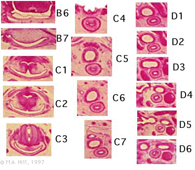
B6:
External auditory meatus (from pharyngeal
groove 1). Meckel's cartilage with
mandible ossifying lateral to it. Tongue
with intrinsic muscles. palatal
processes.Developing teeth.
B7:
Pharynx (compressed dorsoventrally).
Mylohyoid muscle and submandibular gland.
Platysma muscle (R side). Junction of
sigmoid sinus and internal jugular vein on
one side. Vagal ganglia (medial).
Vertebral arteries.
C1:
Pharynx. Carotid neurovascular bundle with
prominent ganglia.
C2:
Pharynx. Carotidneurovascularbundle.
Sternomastoidmuscle.
C3:
Glottic region with cricoid cartilage and
descending process of thyroid cartilage
laterally. Carotidneurovascularbundle.
C4:
Section damaged, but shows thyroid gland
lateral to trachea. Oesophagus.
C5:
Thyroidgland. Trachea. Oesophagus.
Recurrent laryngeal nerve. Common carotid
arteries. Jugular veins. Vagus nerves.
C6,C7:
Jugular veins. In C7 brachiocephalic
vein.
Dl:
Thymus gland.
|