Techniques and Cytology
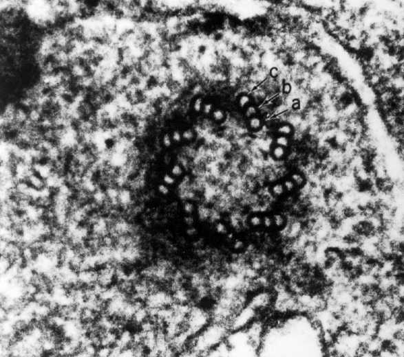
A high magnification electron micrograph showing a centriole in transverse section. Its wall is made up of 9 sets of triplets. Each triplet is composed of 3 subunits: a, b, and c, which form a microtubule.