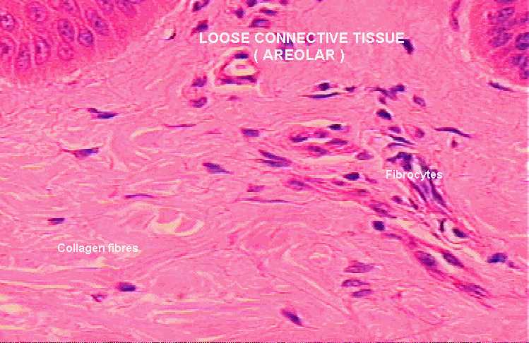
Connective Tissue
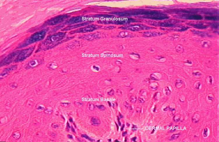
Thick skin
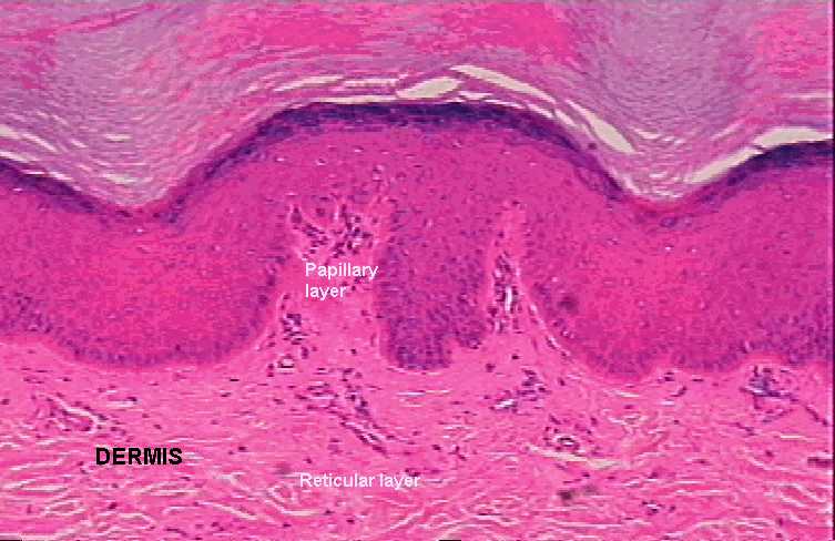
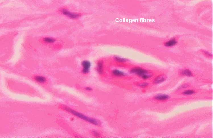
Under X10, locate the papillary layer of the dermis in your slide.(This
layer lies immediately beneath the epidermis). Select a good area of
this layer and move your objective to high-dry(X40). Identify the
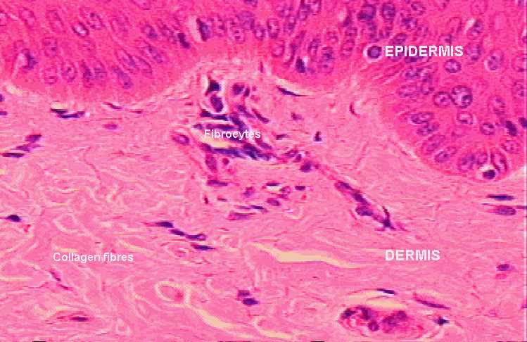
purplish-stained nuclei of fibrocytes. Running between the fibrocytes
are bundles of collagen fibres(pink fibres) which are more prominent as
you move your field down into the reticular layer. Within the papillary
layer(which is composed of loose connective tissue) you may also find
capillaries cut in different planes. Try to identify at least one of
them. Other cell types in the connective tissue are hard to
differentiate. Examine the reticular layer of the dermis. It is composed
of dense irregular connective tissue.
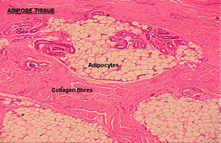
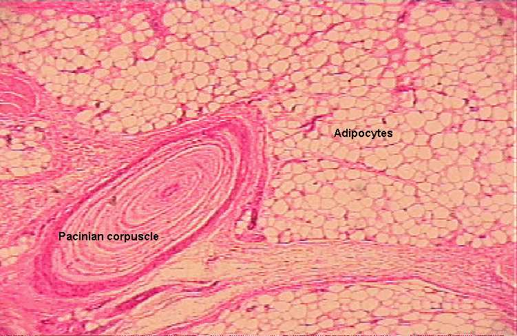
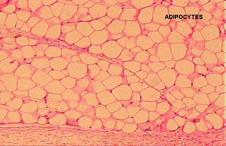
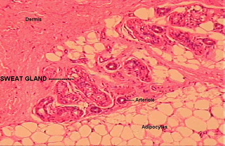
Return your objective to X10 and move your field down to the
subcutaneous layer containing a variable amount of fat(adipose) tissue
and sweat glands. The adipocytes(fat cells) appear empty-looking as
their contents had been washed out during slide preparation. Under X40,
note that the nuclei of the cells are pushed to the periphery of the
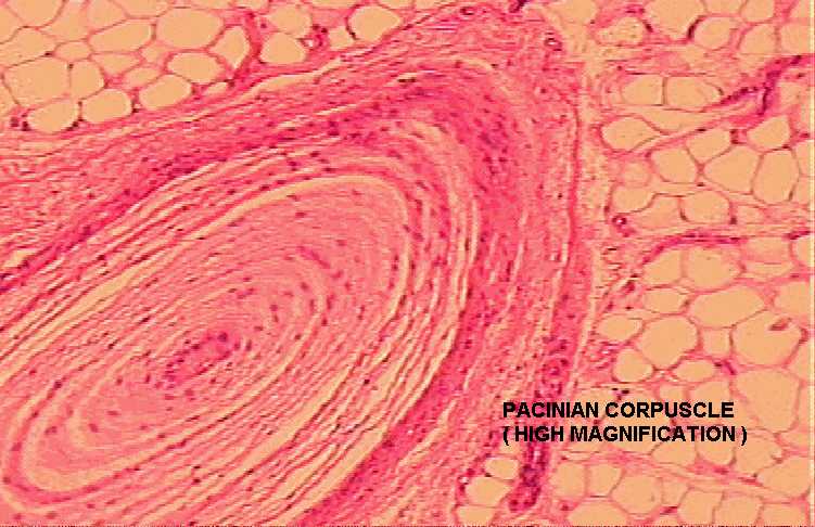
cells. Other structures in the dermis include the sweat glands and
Pacinian corpuscles.