
USC Banco de Imagenes Médicas
USC Banco de Imagenes Médicas
Connective Tissue
Micro 58. LM of loose connective tissue. Note the variety of different connective tissue cells in this field. In the center of the field is a spindle-shaped fibroblast with slender processes and an elongated nucleus.
Micro 59. TEM of a fibroblast. The fibroblast is in the center of the field and contains a dark nucleus. The basal surface and basal lamina of epithelial cells can be seen in the upper left.
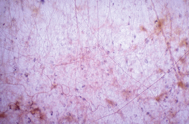
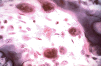
Micro 60. LM of loose connective tissue. Note the collagen and elastic fibers. The thin, taut filaments are elastic fibers which branch and form a network. Collagen fibers are thick and wavy.
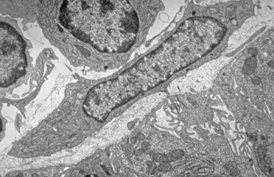
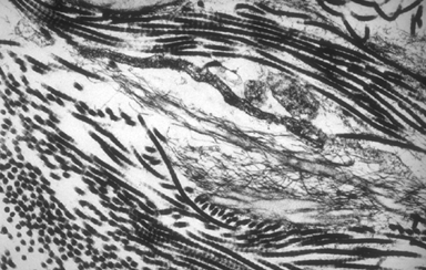
Micro 61. TEM of connective tissue matrix. In the upper right corner are cross and longitudinal sections of collagen fibers. Reticular fibers, in the center, form irregular, fine fibrils.
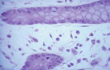
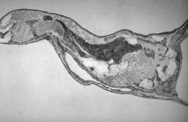
Micro 62. TEM of lung. Simple squamous cells cover the connective tissue core of this alveolar septum. A portion of a fibroblast, the dark cell in the middle, is surrounded by connective tissue fibers and ground substance. Collagenous fibers are seen in cross section, while elastic material appears as a lighter area between the fibroblast and the capillary endothelial cell on the left.
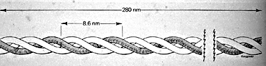
Micro 63. Diagram of a tropocollagen molecule (Junqueira et al., Basic Histology, 1992). The fiber type most commonly associated with connective tissue is type I collagen.
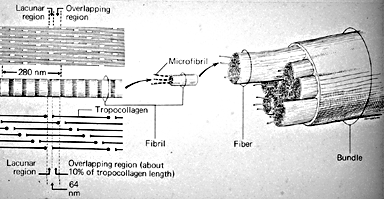
Micro 64 . Diagram showing the assembly of collagen (Junqueira et al., Basic Histology, 1992). This diagram shows the progression from tropocollagen to fibrils, fibers, and bundles.
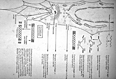
Micro 65. Diagram of the synthesis of collagen by a fibroblast (Junqueira et al., Basic Histology, 1992). The message for each alpha chain is translated on RER and post-translationally modified.
Micro 66. LM of macrophages. Macrophages are large cells derived from blood monocytes. Note the phagocytosed materials.
Micro 67. TEM of a macrophage. Macrophages shows numerous cytoplasmic infoldings, vacuoles and residual bodies.
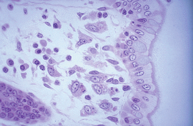
Micro 68. LM of plasma cells. Plasma cells are antibody-producing cells. These cells have an eccentrically-located nucleus, which typically has a cartwheel or clock-face appearance to the heterochromatin, and a well-developed Golgi complex, which is negatively stained in the intensely basophilic cytoplasm.
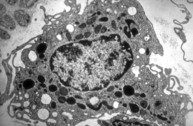
Micro 69. TEM of a plasma cell. This plasma cell is active in protein secretion. Note the euchromatin and heterochromatin in the nucleus and the dilated cisternae of the RER.
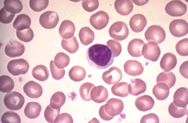
Micro 70. LM of a blood smear. Lymphocytes are small cells with round, densely stained nuclei and a thin rim of bluish basophilic cytoplasm.
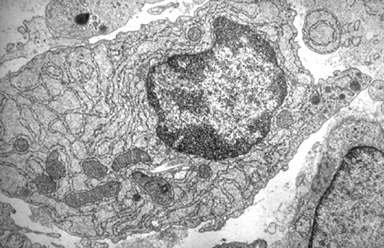
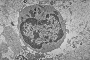
Micro 71. TEM of a lymphocyte. The lymphocyte has a prominent nucleus and a paucity of membranous organelles. There is an abundance of free polyribosomes in the thin cytoplasmic rim. Note the centrioles.
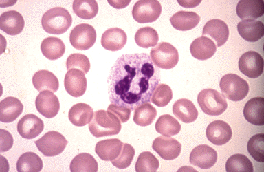
Micro 72. LM of a blood smear. The mature neutrophil has a highly lobulated nucleus, with up to 5 lobes connected by fine strands of nuclear material.
Micro 73. Granulocito con puente y Cuerpo de BARR.- Se le aprecian lisosomas, un nucleo de dos lóbulos unidos por un delgado puente cromatínico; de uno de los lóbulos emerge un pequeńo lobulillo que se interpreta como organizado por un cromosoma X.
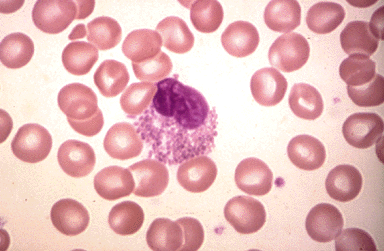
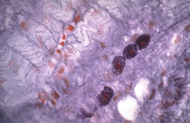
Micro 74. LM of a blood smear. The cytoplasm of eosinophils is packed with large, eosinophilic (dark, pink-staining) specific granules, while the nucleus is often bilobed.
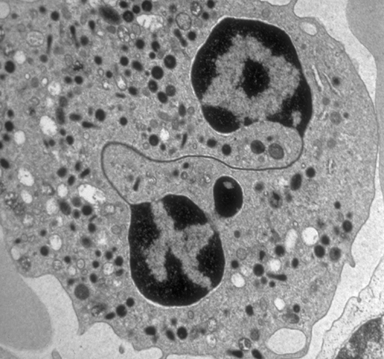
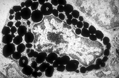
Micro 75. TEM of an eosinophil. The eosinophil has large granules and a bi-lobed nucleus.
Micro 76. LM of a mast cell. Mast cells are found near blood vessels in connective tissue. With suitable staining, they show an extensive cytoplasm, packed with large granules.
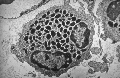
Micro 77. TEM of a mast cell. Mast cells are oval or spherical cells with a heterochromatin-rich nucleus (N); the nucleolus is prominent. The cytoplasm is loaded with many membrane-bounded secretory granules (Gr) which contain histamine and heparin.
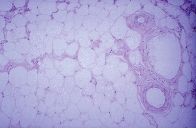
Micro 78. LM of adipocytes (fat cells). Mature white fat cells are large polyhedral cells containing a single central lipid droplet with the nucleus pressed to the cell's periphery and a thin rim of cytoplasm. This is sometimes referred to as a "signet-ring" appearance. Unless special precautions are taken, the lipid dissolves during histological preparation, giving adipose tissue the loose meshwork appearance seen in this Micro.
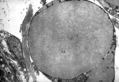
Micro 79. TEM of a fat cell. Note the large lipid droplet (Ld) and the thin-rim of cytoplasm.
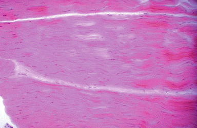
Micro 80. LM of dense regular connective tissue. In dense regular connective tissue large collagen bundles are arranged in the same plane. There are relatively few cellular elements and fibroblasts are the predominant cellular components.
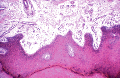
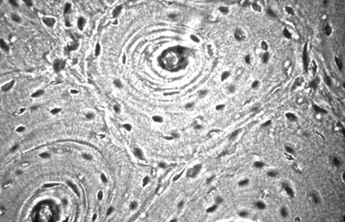
Micro 81. LM of dense irregular connective tissue (skin).
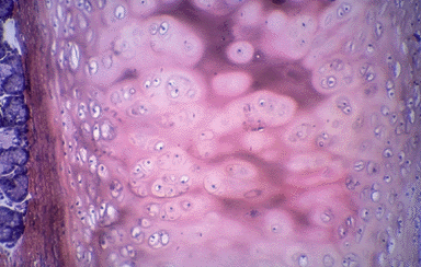
Micro 82. LM of hyaline cartilage. Note the perichondrium at the top. Identify chondroblasts, chondrocytes and lacunae.
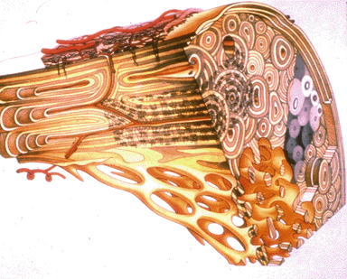
Micro 83. Schematic diagram showing the microstructure of mature bone (Erlandsen and Magney, Human Histology: A Microfiche Atlas).
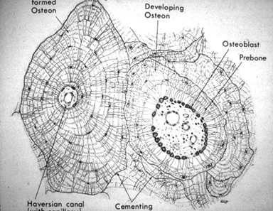
Micro 84. Drawing of osteons (Erlandsen and Magney, Human Histology: A Microfiche Atlas). The diagram shows formation of Haversian systems (osteons) and canaliculi.
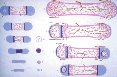
Micro 85. Diagram of endochondral bone formation (Ross and Romrell, Histology, 1989). This process starts with formation of a periosteal bony band (pink) around the diaphysis of a hyaline cartilaginous model (light blue) and continues with endochondral ossification in which hyaline cartilage is destroyed and replaced by bone.
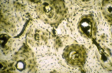
Micro 86. LM of ground bone. Locate osteons, lacunae, canaliculi, osteocytes, Haversian canals and lamellae.
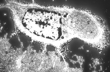
Micro 87. LM of ground bone. Note the osteons in cross-section.
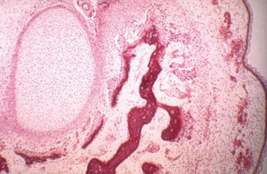
Micro 88. LM of intramembranous bone formation. This image shows intramembranous bone in the pig embryo. The cartilage mass on the right in not directly involved in the bone formation.
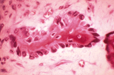
Micro 89. LM of developing intramembranous bone. A bony spicule is located in the center of the field. Osteocytes reside in lacunae completely surrounded by bone matrix, while osteoblasts cover the surface of the spicule.
Micro 90. TEM of newly formed bone. As young, cuboidal osteoblasts (shown here) mature, they become stellate osteocytes with thin processes extending out and making contact with neighboring cells (arrows).
Micro 91. LM of an osteoclast. Osteoclasts are multinucleated cells.