
USC Banco de Imagenes Médicas
USC Banco de Imagenes Médicas
Cell
The Cell
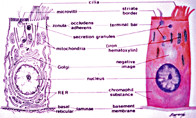
Micro 1. Composite drawings of a typical eukaryotic cell . (Erlandsen and Magney, Human Histology: A Microfiche Atlas). These drawings show characteristic organelles that are detectable by light or electron microscopy.
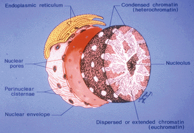
Micro 2. Diagram of the nucleus. (Erlandsen and Magney, Human Histology: A Microfiche Atlas). This diagram shows the major structural features of the nucleus. Note that the nuclear envelope is a double membrane structure with pores.
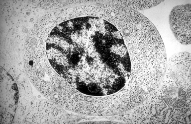
Micro 3. Light micrograph (LM) of spinal cord. (Erlandsen and Magney, Human Histology: A Microfiche Atlas). The large motor neuron in the center of the field has a pale nucleus and prominent nucleolus. The cytoplasm contains basophilic clumps of material containing ribosomes. The clear area around the cell is an example of shrinkage artifact. Note the other, smaller, darkly stained nuclei of various sizes and shapes in the remainder of the field.. Note the row of red blood cells in the capillary in the lower portion of the field.
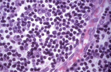
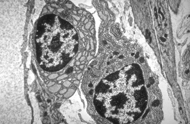
Micro 4. LM of lymph node. (Erlandsen and Magney, Human Histology: A Microfiche Atlas). Note the various sizes, shapes and staining densities of the nuclei in this light micrograph.
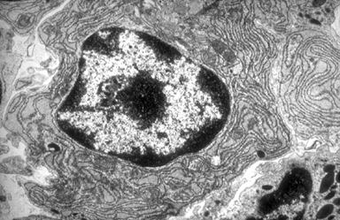
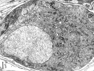
Micro 5. Transmission electron micrograph (TEM) of plasma cell. (Erlandsen and Magney, Human Histology: A Microfiche Atlas). Identify heterochromatin, euchromatin and the nucleolus in this electron micrograph. Note also that the cell shown in this micrograph has abundant rough endoplasmic reticulum.
Micro 6. TEM of parasympathetic neuron (Erlandsen and Magney, Human Histology: A Microfiche Atlas).
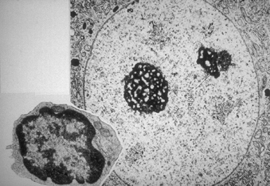
Micro 7. TEMs of two different cell types (Ross and Romrell, Histology, 1989). Note the substructure of the nucleoli evident in the larger micrograph.
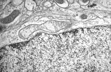
Micro 8. TEM of a portion of a cell (Erlandsen and Magney, Human Histology: A Microfiche Atlas). A segment of a nuclear envelope runs transversely across this electron micrograph. The nucleoplasm is above and the cytoplasm below. Note the double membrane nature of the nuclear envelope and the fact that the envelope is interrupted by nuclear pores.
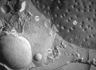
Micro 9. Freeze-fracture of a portion of a cell (Erlandsen and Magney, Human Histology: A Microfiche Atlas). Note the nuclear pores in the nuclear envelope (labelled N). er = endoplasmic reticulum; cm = cytoplasmic matrix.
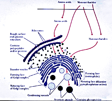
Micro 10. Diagram of synthetic pathway for secreted glycoproteins (Ross and Romrell, Histology, 1989). Note the relationship between rough endoplasmic reticulum (RER) and Golgi apparatus.
Micro 11. TEM of cartilage cells and extracellular matrix (Erlandsen and Magney, Human Histology: A Microfiche Atlas). Note the abundant RER present in these cells.
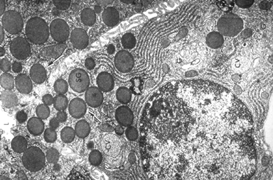
Micro 12. TEM of secretory epithelial cells (Erlandsen and Magney, Human Histology: A Microfiche Atlas). Note, the numerous large secretory vesicles (SG) in this cell.
Micro 13. TEM of a liver cell (Erlandsen and Magney, Human Histology: A Microfiche Atlas). This high magnification electron micrograph shows the association of ribosomes with the membranous cisternae that form the RER. Note also the presence of mitochondria and glycogen deposits in this field.
Micro 14. TEM of connective tissue cells (Erlandsen and Magney, Human Histology: A Microfiche Atlas). Note the dilated cisternae of the RER in the cell on the left.
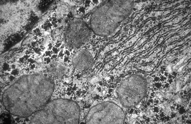
Micro 15. TEM of portion of neuron. Identify the Golgi apparatus in this electron micrograph. Note the coated vesicles that are present.
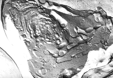
Micro 16. Freeze-fracture of portion of a cell. Identify the Golgi in this freeze-fracture image.
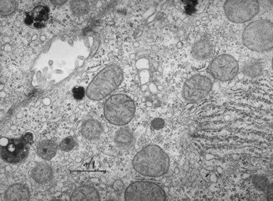
Micro 17. TEM of portion of a cell (Erlandsen and Magney, Human Histology: A Microfiche Atlas). Circular profiles of tubulo-vesicular smooth endoplasmic reticulum (SER) are shown in this micrograph (along with some RER, mitochondria and dense-cored peroxisomes).
Micro 18. TEM of developing RBC (Erlandsen and Magney, Human Histology: A Microfiche Atlas). Note lack of endoplasmic reticulum in this cell.
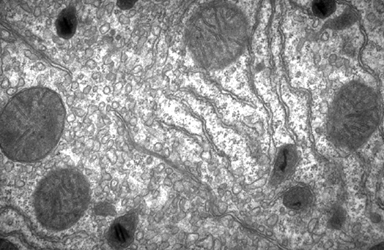
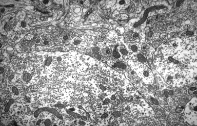
Micro 19. TEM of the CNS. Many free polyribosomes and profiles of RER can be identified in the cell body in the upper right of this electron micrograph.
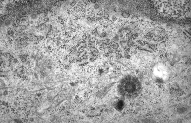
Micro 20. TEM of portion of cytoplasm and tangential cut of nucleus (Erlandsen and Magney, Human Histology: A Microfiche Atlas). Ribosomes in the polysome configuration can be identified in this high magnification electron micrograph. Note also the centriole in the lower right and nuclear pores at the top.
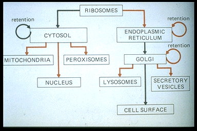
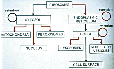
Micro 21. Diagram of synthetic pathways. Note the differing fates of proteins synthesized by free polyribosomes as opposed to ribosomes that are associated with the endoplasmic reticulum.
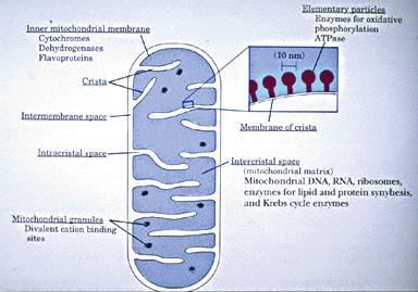
Micro 22. Blank Micro 23. Diagram of a mitochondrion (Ross and Romrell, Histology, 1989). Note the double membrane and the fact that the inner membrane is thrown into folds called cristae.
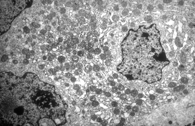
Micro 24. Low magnification TEM (Erlandsen and Magney, Human Histology: A Microfiche Atlas). Mitochondrial morphology varies based on functional state, cell type and, of course, plane of section. Note the round mitochondrial profiles in this electron micrograph.
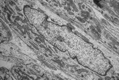
Micro 25. Low magnification TEM (Erlandsen and Magney, Human Histology: A Microfiche Atlas). Note the elongated mitochondria shown in this micrograph.
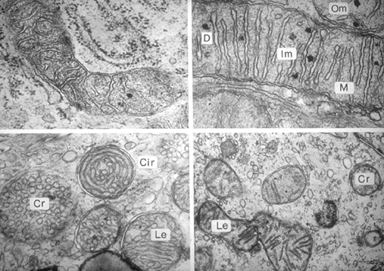
Micro 26. High magnification TEM of mitochondria (Erlandsen and Magney, Human Histology: A Microfiche Atlas). Cristae vary in morphology, as seen in these micrographs of four different cell types. Cr, Cir & Le = cristae in different planes of section; D = dense granule; Im = inner membrane; Om = outer membrane; M = matrix.
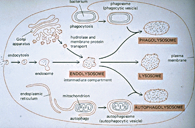
Micro 27. Diagram of degradation pathways. Utilize this diagram to understand the similarities and differences in the various pathways of the lysosomal system.
Micro 28. TEM of two adjacent liver cells. Note the darkly-stained lysosomes containing partially digested material.