Part 8: Cardiovascular System
To search for specific words try the "Find" and "Find Again" commands on your browser.
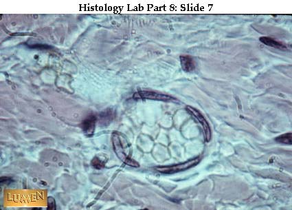
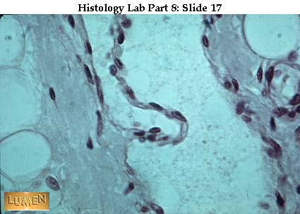
This capillary running through embryonic mesenchyme has a wall consisting solely of a single layer of endothelium. Notice that the lumen of the vessel is only slightly larger than the diameter of the r.b.c.'s within.
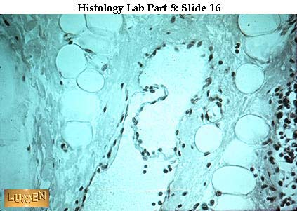
This sinusoid, like a capillary, has only an endothelial wall, but its lumen is characteristically considerably wider. Also, in some locations in the body (such as bone marrow, liver, and spleen) the endothelial cells of sinusoids are rather loosely joined together, thus permitting passage of blood cells between them.
In the middle of the field is a sinusoid (filled with orange-colored r.b.c.'s) in the marrow cavity of spongy bone. (The larger empty circles are fat cells.)
A capillary lying in the endomysium between skeletal muscle fibers. This one shows very dark endothelial nuclei and has 3 pink r.b.c.'s lined up in a row inside.
EM of different capillary endothelia.
- A) continuous, endothelium in capillary of cardiac muscle.
- B) continuous, thin endothelium of lung capillary with a thin layer of lung epithelium underlying it at (6).
- C) fenestrated endothelium of capillary of kidney.
Note: basal lamina (3) under each one. Also pinocytotic vesicles (5), fenestrations (10), and endothelial cytoplasm (2). In each picture, (1) = space of capillary lumen; (4) = connective tissue space outside. (Don't worry about #8 yet!)
Diagram of routes of transport across capillary or sinusoidal endothelial cells. (Notice that we now also consider discontinuous endothelium as well as the types you saw on the previous slide.)
- A) capillary lumen
- B) basal lamina
- C) pericapillary space (i.e., connective tissue ground substance)
- D) tight junction (fused outer leaflets)
- E) gap junction (2-4 nm or 20-40 A intercellular space)
- Fl-3) adjoining portions of separate endothelial cells that contact each other. Their ordinary intercellular distance is 150-200 A (15-20 nm).
"Spikes" on the outer leaflet of membrane = glycocalyx layer.
- 1 = diffusion through plasma membrane
- 2 = pinocytotic vesicles
- 3 = tight junction (impenetrable)
- 4 = gap junction (penetrable by small molecules)
- 5 = fenestration with closing membrane (flow allowed selectively)
- 6 = open fenestration within the cytoplasm of a given endothelial cell (easy passage of fluid and molecules)
- 7 = hole, discontinuity between endothelial cells (note that basal lamina is also gone)
Endothelium (simple squamous epithelium) lines the lumen of all blood and lymph vessels, as well as the heart. Here endothelial nuclei are seen ringing a venule (so identified because there is only a thin coat of connective tissue outside the endothelium, the lumen is too large to be a capillary, and because sinusoids don't occur in ordinary conn
ective tissue areas like this.)
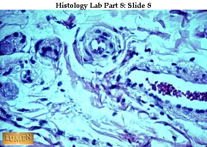
The smallest arterioles have a single layer of smooth muscle outside the endothelium, and a lumen hardly wider than a capillary. Toward the upper center of this field is a round, cross-cut arteriole with just one or two layers of muscle in the media. To the right is the more irregular, wider shape of a venule with only a thin adventitial wall.
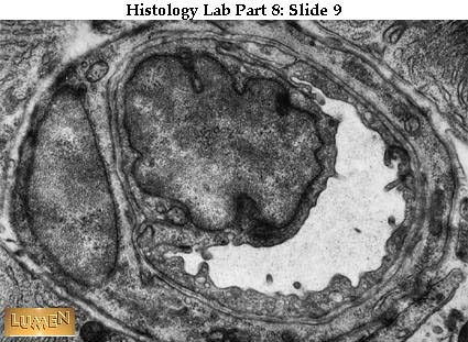
EM of capillary. The inner nucleus and ring of cytoplasm is the endothelial cell around the lumen. The outer nucleus and ring of cytoplasm is a single pericyte wrapped around the vessel. Notice the thin gray line of basal lamina between the cytoplasm of the two cells and shared by both cells. There is also a basal lamina along the outer surfac
e of the pericyte as well.
Small blood vessels, with 3-layered walls:
- a = small lymphatic with endothelial lining but no other organized wall.
- b = small vein (very thin muscle; pale c.t.).
- c = small artery (pink muscle; yellow c.t.).
These vessels are often found running together in the c.t. coats of body organs.
A small artery cut longitudinally. Notice the circular arrangement of smooth muscle cells cut tangentially at the left end. For most of the vessel, the muscle is cross-cut, looking almost like an epithelium. The real epithelium, however, is the simple squamous endothelium immediately lining the lumen, with thin, flat nuclei oriented longitudina
lly along the vessel.
A medium-sized (muscular) artery, showing the typical 3 wall layers:
- (1) tunica intima, consisting of endothelium lining the lumen, and a small amount of areolar c.t., a black and wavy inner elastic membrane divides this layer from the tunica media. (Only the elastic membrane shows up at this magnification.)
- (2) tunica media = smooth muscle (bluish color here) and some elastic fibers (black).
- (3) tunica adventitia = dense irregular c.t. (pink collagenic fibers, black elastic fibers). There is fat outside the adventitia.
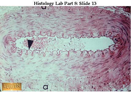
Another medium-sized, muscular artery (also called a distributing artery). This is typical of the arteries you dissected in the arm; it usually runs with a vein and nerve. There is a characteristic inner elastic membrane (dark pink with arrow pointing to it) and a heavy circular muscle in tunica media. Note that a = adventitia.
EM photo of inner elastic membrane (white band). Endothelial cells are bunched up because the wall is contracted.
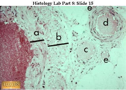
Medium-sized vein with a much less compact muscle layer than you saw in the preceding arteries. The tunica media is indicated by bar "a". Bar "b" = adventitia, which is at least as wide as the media, and often even wider. There is no evident inner elastic membrane. (Blood in the lumen stains red here.) To the right, compare sizes and walls of
one small artery (d) and two very small veins (c) and (e).
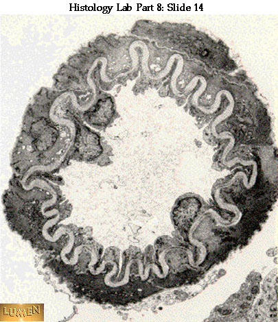
Lymphatic vessel with a c.t. wall even thinner than a vein. There is a cut leaflet of a valve across the lumen. Material in the lumen contains no r.b.c.'s, mostly just structureless lymph and some lymphocytes. There are some fat cells and lymphocytes in the surrounding connective tissue.
Higher power of valve made of a core of fine c.t. with endothelium covering both surfaces. Valves in veins are constructed similarly.
Low power view of wall of aorta, an elastic artery:
- a = intima (endothelial lining plus thin layer of underlying c.t.).
- b = media (alternating layers of elastic membranes and smooth muscle, bound together by areolar c.t.) This is by far the thickest layer.
- c = adventitia (fairly dense c.t. carrying small blood vessels, the vasa vasorum).
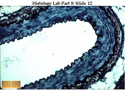
Detail of inner portion of aortic wall: "a" bar = depth of tunica intima (next to lumen). In the media there are many layers of wavy, dark-stained elastic membranes, alternating with the paler pink smooth muscle and areolar c.t. (Note that these are elastic membranes, not just fibers. Think of many layers of rubber sheets enwrapping the vessel,
and you have cut across them and are looking at the cut edges. These sheets are fenestrated; i.e., they have holes in them, thus allowing passage of nutrients diffusing from the blood in the aortic lumen out into the tissues of the wall.) Arrows = nuclei of smooth muscle cells.
Detail of outer portion of aortic wall, showing blood vessels (vasa vasorum) in the adventitia. These vessels bring nutrients only to the outer one-third or so of the vessel wall.
- a = small nerve (very pale here).
- b = small vein - very little muscle, mostly pale c.t..
- c = small artery - considerable muscle, some c.t. (elastic fibers specifically stained and mixed with collagenic fibers). The c.t. adventitia just merges with the surrounding packing c.t..
Heart
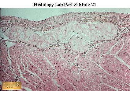
Inner surface of the heart, with pale, large Purkinje fibers lying in the subendocardial layer. Endocardium (or intima) is above. The beginning of the myocardium (media, cardiac muscle) is below.
The heart wall, like blood vessels in general, has three main layers, though they are not called intima, media, and adventitia. As in vessels, however, the innermost and outermost layers are primarily connective tissue; the middle one is muscle --- in this case, cardiac muscle. From left to right, then, in this picture of ventricle wall, there i
s first a very thin endocardium, which consists primarily of an endothelial lining and a very small amount of connective tissue underneath it. The muscle layer, or myocardium is next and is by far the thickest layer and constitutes the bulk of the heart. To the far right is the epicardium, which contains considerable fat. In gross anatomy the e
picardium is called the visceral layer of the serous pericardium; it has an outermost lining layer of mesothelium.
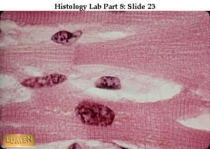
A high magnification reminder of the appearance of cardiac muscle cut longitudinally, with central nucleus, branching fibers, and cross-striations. Muscle fibers spiral around the heart in all directions and can thus exert the necessary squeezing action as the heart contracts. Remember that these muscle cells are attached end to end by junctions
at the intercalated disc. Axon terminals of autonomic neurons innervate some of the muscle cells, and the stimulus is spread to neighboring muscle cells by the intercalated discs and by gap junctions along the side walls of the cells.
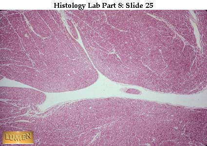
Cardiac muscle in cross-section. Note also the many cross cut capillaries in the connective tissue endomysium between muscle fibers. As you might expect from the constant work the heart performs, it is a highly vascularized organ. Capillaries in this (or any) muscle have endothelium that is continuous and non-fenestrated.
The surface of the ventricular lumen is very irregular because of the presence of papillary muscles in the wall. These irregularities are, of course, lined with endothelium.
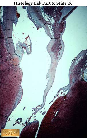
Low power of a Mallory-stained heart, showing two channels (above) that are continuous with the lumen of the left ventricle (below). The left-hand channel is the aorta, with some blue connective tissue in its wall. There is also one cusp of the semilunar valve, with its blue core of dense collagen. Remember that valves are lined over their enti
re surface by endothelium which is continuous with aortic endothelium above and the ventricular endothelium below. To the right in this picture is the atrioventricular channel, with chordae tendinae extending down from the mitral valve and attaching to the papillary muscles of the ventricle. Like valves, the chordae tendinae are also composed of
dense collagenous connective tissue covered by an endothelial lining.


