GRAM-POSITIVE BACILLI<BR>(NON-SPORE-FORMING)
GRAM-POSITIVE BACILLI
(NON-SPORE-FORMING)
ACTINOMYCES
(under the microscope)
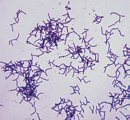 Actinomycetes are fungus-like bacteria that form filamentous branches. These Gram-positive obligate anaerobes
are known to reside in the mouth and in the intestinal tract. Pathogenic proliferation of the
organisms, which is usually a result of trauma to the region of infection, can lead to actinomycosis.
The patient will form abscesses and swelling at the site of infection. A diagnosis can be made upon
microscopic examination of pus. The fluid will have a granular texture which is caused by sulfur
granules. These sulfur granuules are actually composed of the bacterium and its waste. The species of
Actinomyces which is most commonly associated with actinomycosis is A. israelii, but several
other bacteria in this genus are capable of causing the disease as well.
Actinomycosis can often be treated with large amounts of penicillin or tetracycline.
Actinomycetes are fungus-like bacteria that form filamentous branches. These Gram-positive obligate anaerobes
are known to reside in the mouth and in the intestinal tract. Pathogenic proliferation of the
organisms, which is usually a result of trauma to the region of infection, can lead to actinomycosis.
The patient will form abscesses and swelling at the site of infection. A diagnosis can be made upon
microscopic examination of pus. The fluid will have a granular texture which is caused by sulfur
granules. These sulfur granuules are actually composed of the bacterium and its waste. The species of
Actinomyces which is most commonly associated with actinomycosis is A. israelii, but several
other bacteria in this genus are capable of causing the disease as well.
Actinomycosis can often be treated with large amounts of penicillin or tetracycline.
LABORATORY INDICATIONS:
- Indole -
- Catalase -
- Lipase -
- DNase -
BIFIDOBACTERIUM
(under the microscope)
 Members of the genus Bifidobacterium are anaerobic, Gram-positive bacilli that is rarely associated with
infection. The only pathogenic species of this genus is Bifidobacterium dentium, a normal inhabitant
of the gut flora. Under the microscope, these bacteria appear to be bone shaped, which makes them easy to
identify. As obligate anaerobes, they require a very low oxygen tension to survive and to achieve moderate
growth.
Members of the genus Bifidobacterium are anaerobic, Gram-positive bacilli that is rarely associated with
infection. The only pathogenic species of this genus is Bifidobacterium dentium, a normal inhabitant
of the gut flora. Under the microscope, these bacteria appear to be bone shaped, which makes them easy to
identify. As obligate anaerobes, they require a very low oxygen tension to survive and to achieve moderate
growth.
LABORATORY INDICATIONS:
- Non-motile
- Catalase -
- Forms branching filaments
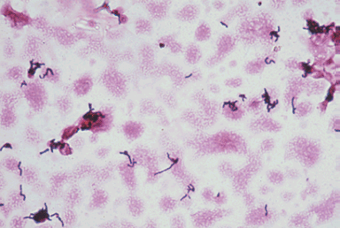
EUBACTERIUM
(under the microscope)
 Eubacterium are normal flora of
the intestinal tract and may cause opportunistic infections. E. lentum, the most isolated
species, has been linked to endocarditis and some wound infections. Biochemical testing can distinguish
Eubacterium from the other Gram-positive, anaerobic rods. Because Eubacterium species are negative
for many tests, results may be somewhat ambiguous. It is important to know, however, that these bacteria tend
to form clumps under microscopic observation.
Eubacterium are normal flora of
the intestinal tract and may cause opportunistic infections. E. lentum, the most isolated
species, has been linked to endocarditis and some wound infections. Biochemical testing can distinguish
Eubacterium from the other Gram-positive, anaerobic rods. Because Eubacterium species are negative
for many tests, results may be somewhat ambiguous. It is important to know, however, that these bacteria tend
to form clumps under microscopic observation.
LABORATORY INDICATIONS:
- Indole -
- Catalase -
- Hydrogen sulfide -
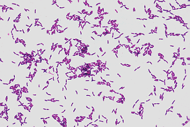
PROPIONIBACTERIUM
(under the microscope)
 Propionibacterium species are some of the most common Gram-positive anaerobes that are isolated in the
laboratory. One particular species, P. acnes, is a usually harmless microbe that has pathogenic
potential. It has been linked to certain cases of endocarditis, wound infections, and abscesses. Ironically,
it can infect acne sites on the skin but it does not cause them. Under the microscope,
Propionibacterium clump up and may show a slight tendency to branch. Also, they show uneven staining
patterns following a Gram-stain procedure. Colonies grow best in an anaerobic or microaerophilic environment
using blood agar.
Propionibacterium species are some of the most common Gram-positive anaerobes that are isolated in the
laboratory. One particular species, P. acnes, is a usually harmless microbe that has pathogenic
potential. It has been linked to certain cases of endocarditis, wound infections, and abscesses. Ironically,
it can infect acne sites on the skin but it does not cause them. Under the microscope,
Propionibacterium clump up and may show a slight tendency to branch. Also, they show uneven staining
patterns following a Gram-stain procedure. Colonies grow best in an anaerobic or microaerophilic environment
using blood agar.
LABORATORY INDICATIONS (P. acnes):
- Indole +
- Catalase +
- Glucose fermentation
- Gelatin hydrolysis


Copyright © 1995 University of Texas - Houston Medical School, DPALM MEDIC, All rights reserved.