Tay-Sachs disease
Click picture to enlarge. Close window to return
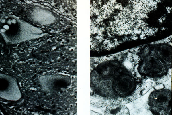
These are ganglion cells associated with Tay-Sachs disease. A is a light microscopic view of a large neuron at top with obvious lipid vacuolation with karyolysis and granularity of nucleus. B is an electron micrograph of a portion of a neuron showing prominent lysosomes with whorled configurations.