Inflammatory edema - postcapillary venule as the targe
Click picture to enlarge. Close window to return
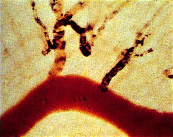
This is a photomicrograph of living tissue to demonstrate which vessel(s) become leaky after the experimental injection of histamine. The animal was first injected with carbon particles, after which histamine was given. After several minutes needed to allow excess carbon particles to clear from the circulation, only one kind of vessel had carbon particles in its wall: the postcapillary venule. You can clearly see that here, where the red, relatively large diameter venule at the bottom has no labeling of its walls; the capillary bed in the background has little or no labeling; but the connecting, postcapillary venules have taken up significant amounts of the label. Note how sharp the cut-off is: you can actually see the carbon-labeling stop, making a sort of footprint, where the postcapillary venules join the larger caliber venule.