Concentric layers of a granuloma
Click picture to enlarge. Close window to return
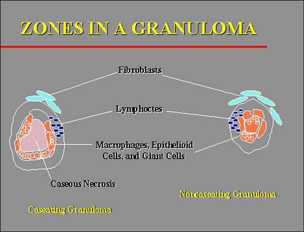
Granulomas are generally divided into zones, as is represented here. This cartoon is simplistic, because there are not as sharp lines of demarcation as are drawn here; cells from each zone usually overlap into adjacent zones, so that the lines of demarcation are blurred. Remember, that this structure forms as an attempt either to eliminate the inciting cause or, if that fails, to wall it off. The center of the lesion depicted on the left contains amorphous debris that is characteristic of caseous necrosis. It is, therefore, a caseating granuloma. This central zone is then surrounded by one that contains epithelioid cells, multinucleated giant cells, and macrophages (from which the first two cell types are formed). Around them is usually a zone of lymphocytes, especially if the granuloma has been formed in conjunction with an immune response against the inciting antigen (a so-called "immune" granuloma, compared to one that is formed against a relatively inert particle, like silicon). To have the lymphocytes in this zone makes good sense, because it is they that secrete the mediators that activate and alter the macrophages and macrophage-derived cells that are located centrally. The outer zone is one that contains fibroblasts, which are there to help to wall off the lesion with fibrous connective tissue. The cartoon on the right has the same zones, with the exception that there is no zone of central necrosis. It is, therefore, referred to as a noncaseating granuloma. With this orientation, you should now be able to recognize the concentric components of the real granulomas that are shown in the following two photomicrographs