Concentric layers of a granuloma
Click picture to enlarge. Close window to return
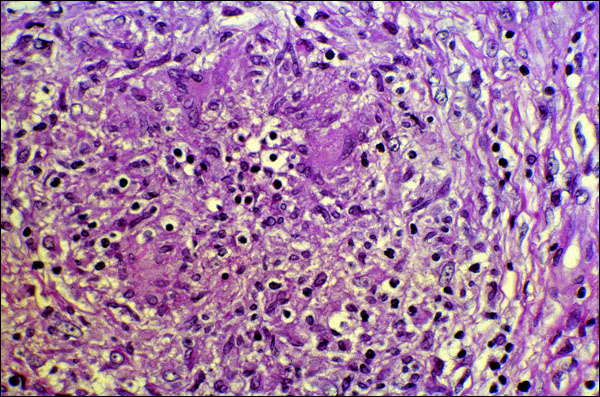
epithelioid cells, multinucleated giant cells, and macrophages (from which the first two cell types are formed). Again, It is emphasized that there are no sharp lines of demarcation between the various zones -- they tend to grade into each other. Giant cells are more easily seen in this granuloma, e.g., there is a representative one at approximately 2:30 o'clock, just away from the center. Note the number of pyknotic nuclei that can be seen -- the dense, homogeneous, almost black structures that are surrounded by clear rings (shrinkage artifact associated with fixation) throughout the field. Epithelioid cells can be seen outside the ring of giant cells, and a concentric ring of fibroblasts can be seen at the right of the picture. Lymphocytes are scattered throughout several of the layers (just slightly larger and less dense than the pyknotic nuclei). In some kinds of granulomatous inflammation, granulomas are very poorly defined. For example, the tuberculoid form of leprosy has distinct granulomas associated with it, while those associated with the lepromatous form -- the kind of leprosy that is poorly controlled -- are generally poorly formed.