Chronic inflammation
Click picture to enlarge. Close window to return
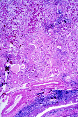
This is a low-power photomicrograph of a kidney that has been damaged extensively by chronic pyelonephritis. The pelvis, lined by epithelium, is the open cleft at the bottom of the field. At higher power we would see that the basophilic band above the pelvis is composed of chronic inflammatory cells, i.e., lymphocytes, macrophages and plasma cells. Similar, but less prominent, aggregations of chronic inflammatory cells can be seen elsewhere in the tissue. Fibrosis is a characteristic of chronic inflammation. As maturation of fibrous connective tissue proceeds, it contracts, often constricting parenchymal tissue. You can see that outcome here, because tubules have been blocked, resulting in their becoming distended to varying degrees with pink, protein-containing fluid. By comparing this section with that of a normal kidney, e.g., a section of the renal medulla in a histology text, you will also see that the architecture has become distorted, due to parenchymal loss and fibrosis. It's not hard to imagine that this kidney was dysfunctional.