Tissue injury by inflammatory cells
Click picture to enlarge. Close window to return
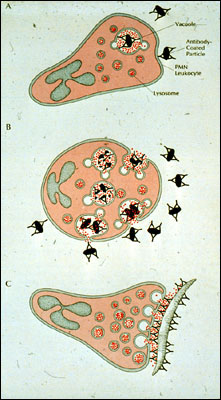
(The captions in the upper right corner, from top to bottom, read: "vacuole","antibody-coated particle", "PMN leukocyte", and "lysosome"). The upper panel in this figure shows the ideal scenario for phagocytosis of a bacterium opsonized with antibody: there is no loss of hydrolytic enzymes to the extracellular compartment. The middle panel shows what can happen when the phagocytic stimulus is so great that it persistently stimulates the neutrophil. Under these conditions (regurgitation while feeding), lysosomes begin fusing with phagocytic vacuoles before they are fully closed, resulting in extracellular loss of lysosomal enzymes and reactive oxygen intermediates. The result is indiscriminate injury of adjacent tissue. The lower panel shows what happens when a particle is too large for a phagocyte to ingest. It adheres to the surface and redistributes its lysosomes to that side of the cell. The lysosomes fuse with the plasma membrane, thereby ejecting their contents onto the non-ingestible surface. This process is often referred to as "frustrated phagocytosis", and is a major cause of tissue injury in certain antibody-based autoimmune diseases