Leukocyte exudation
Click picture to enlarge. Close window to return
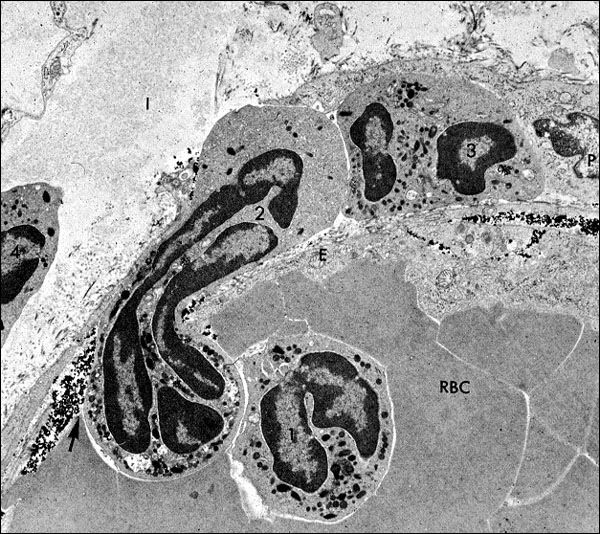
This is an electronmicrograph of a neutrophil caught in the act of exiting a vessel. This process is dependent on the leukocyte locally breaking down the vessel's basement membrane, by directed secretion of hydrolytic enzymes onto the surface of the BM. Erythrocytes in the vessel's lumen are labeled, "RBC", and endothelial cytoplasm that lines the vessel on either side of the emerging neutrophil is labeled, "E". A neutrophil, labeled "1", is still in the lumen. The emerging neutrophil is labeled "2"; one that has reached the outer limits of the vessel wall is labeled "3", next to a pericyte (P) at the right edge of the frame; and one that is free in the extravascular space is labeled "4", at the left edge of the micrograph. Carbon particles are present in the wall of the vessel, under the endothelium, giving proof that this postcapillary venule was in a state of increased permeability (due to endothelial cell contraction caused by inflammatory mediators, like histamine).