Suppurative inflammation
Click picture to enlarge. Close window to return
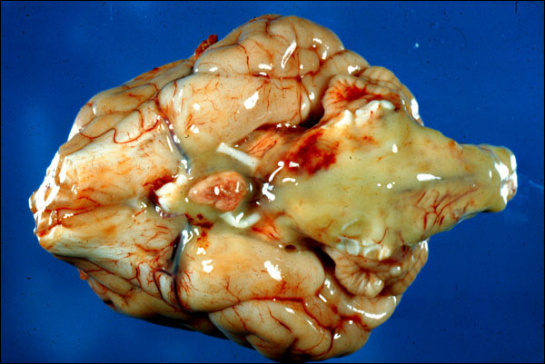
This is the brain from a year-old mongrel dog that had suppurative osteomyelitis of vertebral body, C1. It has led, by extension, to suppurative meningitis, which is depicted here. This could just as easily be the brain of a human being, because the disease and the process of extension of an inflammatory process into adjacent tissue are the same in each species. The pituitary gland can be seen at the base of the brain, slightly to the left of center. Surrounding the pituitary, and extending posteriorly from it, there is yellow-white, creamy looking material, which is pus. This dog (or a patient with the comparable process) would have had neutrophils and increased amounts of protein in its cerebrospinal fluid.