Exudate
Click picture to enlarge. Close window to return
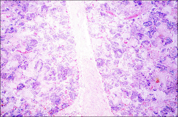
Your text tells you that an exudate differs from a transudate, in that the former contains more protein (specific gravity >1.015) and often contains leukocytes. This photomicrograph of an acutely inflamed lung dipicts clearly what an exudate is. Note that the alveoli, as well as the interstitial tissue that separates the two lobules, are filled by pink material. That is the way that protein looks in a routine slide stained with hematoxylin and eosin dyes (H & E, the routine stain used to study tissues microscopically). The protein was contained in edema fluid, which must have been plentiful, because the alveoli are distended, and the two lobules are uncharacteristically separated from each other, i.e., the interstitial space is widened. In this field, you can additionally see that leukocytes are present in this exudate, although they cannot be specifically identified as to type at this low power. They form the basophilic aggregates that are scattered widely in the alveolar spaces. Another clue that this is acute pneumonia is the hyperemia that is seen throughout this section.