Carcinoma of the prostate, histology, high power
Click picture to enlarge. Close window to return
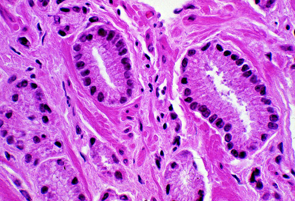
Here the structure of the neoplastic glands is seen to better advantage. Note that no myoepithelial cell layer can be seen near the basement membrane (compare to the slide of benign prostatic hypertrophy). Also, "back to back" gland formation, without intervening stroma, is seen best in the lower left hand corner of the photomicrograph.