Malakoplakia, histology and electron microscopy
Click picture to enlarge. Close window to return
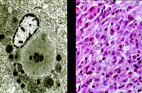
The chronic inflammatory infiltrate in the wall of the urinary bladder consists of macrophages that have well developed eosinophilic cytoplasm. Many of the macrophages contain round, bluish cytoplasmic inclusions. Some of these Michaelis-Gutmann (MG) bodies appear targetoid i.e. have a centrally located dot, surrounded by a clear halo. By electron microscopy, the nascent MG bodies are giant lysosomes with a calcified center surrounded with amorphous proteinacedus material.