Celiac disease, histology
Click picture to enlarge. Close window to return
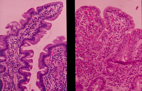
The left portion of the photograph shows normal small intestinal villi. The right part of the picture shows loss of villi and elongation of the crypts typical of celiac disease.