Adenocarcinoma arising in Barrett esophagus, histology
Click picture to enlarge. Close window to return
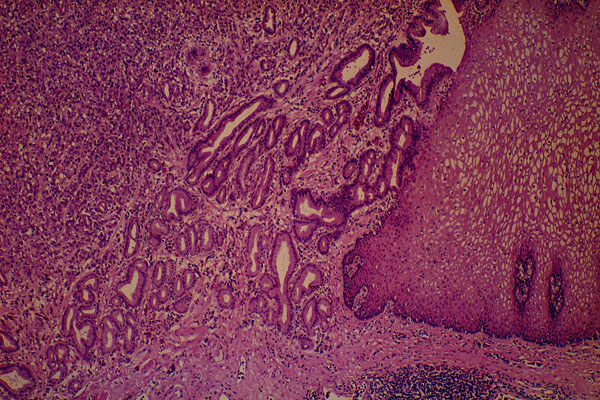
The right portion of the picture shows normal squamous epithelium. The left part of the photograph is occupied by a tumor. Tumor cells are arranged into glands and solid strands.