Blastomyces dermatitidis, H&E stain.
Click picture to enlarge. Close window to return
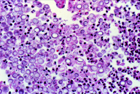
This slide shows neutrophils, histiocytes and numerous fungal organisms which appear as round, gray structures. The organisms are well visualized without silver stains because of the thick cell wall.