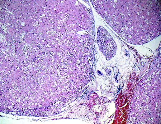
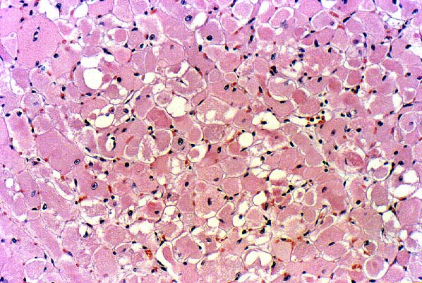
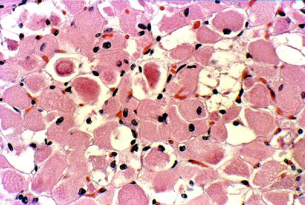
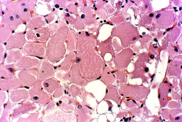
MICROSCOPIC DESCRIPTION:
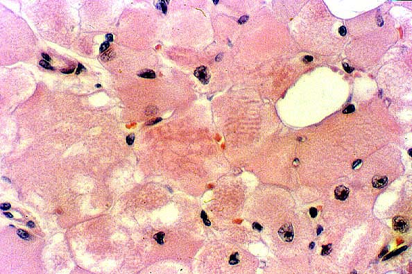
The tumor is well-circumscribed, but unencapsulated and composed of multiple well-demarcated lobules (micro 1). The tumor cells are polygonal, uniform and densely packed with abundant eosinophilic cytoplasm (micro 2). Cytoplasmic vacuolization is evident (micro 3) with both centrally and peripherally located monomorphic nuclei (micro 4). Mild variation in tumor cell size and shape can be seen along with occasional well-oriented cytoplasmic cross-striations (micro 5).